|
|
HOW TECH TICKS |
|
|
|
|
|
|
|
x 6,000
|
|
|
|
|
x 10,000
|
|
|
|
|
|
|
by JESSE TUEL |
|
x 300,000 |
|
Photos by Jim Stroup
Microscopic images courtesy of Mitsu Murayama |
|
|
|
|
|
|
|
|
| The transmission electron microscope |
|
|
Using a multimillion-dollar transmission electron microscope, Virginia Tech researchers captured 130 images of a mineral called schwertmannite, magnified about 6,000 times. Stitching the images together produces this three-dimensional view.
|
|
|
|
|
|
|
|
|
|
The mineral sample was pulled from a river adjacent to ancient mining sites in Spain. Magnifying the images further, to about 300,000 times, researchers can inspect individual atoms and determine whether arsenic, which tends to bind itself to the mineral and is thus ferried to the ocean, is encapsulated in the mineral's interior or, more dangerously, exposed to the water on the exterior.
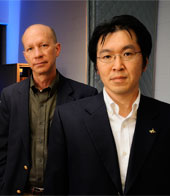
The images are courtesy of University Distinguished Professor of Geosciences Michael Hochella Jr. and Mitsu Murayama, associate professor of materials science.
|
|
|
|
In Virginia Tech's Corporate Research Center, on a building site selected for its seismic passiveness, in a cubed room whose temperature won't deviate more than a 10th of a degree, behind ceiling-high curtains that block out the most benign air flow, a multimillion-dollar transmission electron microscope (TEM) sees individual atoms that are much less than one nanometer in size. (A human hair is 80,000 nanometers wide.) Such precision requires the utmost stillness.
Acquired about five years ago, the microscope was first housed in Derring Hall. The instrument is so sensitive that researchers could tell if someone was walking in the building or if a car was being driven in the parking lot.
|
|
|
|
|
|
Michael Hochella Jr. '75, '77 and Mitsu Murayama
|
|
|
|
|
The TEM is now housed in the Nanoscale Characterization and Fabrication Laboratory (NCFL), specifically designed for such delicate devices. "If Mitsu [Murayama, an associate professor of materials science] is taking an image at atomic resolution, we don't even talk in this room," said University Distinguished Professor of Geosciences Michael Hochella Jr. (geological sciences '75, M.S. '77).
In the span of one second, approximately 10 billion electrons are fired downward, screaming at nearly the speed of light through a column so highly evacuated of air that the cylinder's atmospheric properties are close to that of outer space. Magnetic lenses focus the electron beam's trajectory toward where the sample rests below. Whereas a light microscope bends light rays to produce an image, the TEM capitalizes on the incredibly small wavelength of electrons to detect the electrons' reaction when they pass through the sample.
The results are stunning. By stitching together 130 TEM images of a mineral called schwertmannite, Murayama has produced a three-dimensional view of a mineral sample pulled from a river adjacent to ancient mining sites in Spain. Zooming into the 20-nanometers-wide mineral whiskers, the microscope reveals layers of atoms, like beaded necklaces stacked on the computer screen. With this atomic-level view, researchers can examine whether arsenic, which tends to bind itself to the mineral and is thus ferried to the ocean, is encapsulated in the mineral's interior or, more dangerously, exposed to the water on the exterior.
"It's these details that tell us how these materials behave in nature and whether they're going to be dangerous," said Hochella, who was the first in the emerging field of nano-bio-geochemistry to use an instrument like the TEM to study surface properties at the atomic level. From arsenic in Spanish rivers to acid-mine drainage in Germany to toxic metals in Montana's Clark Fork River, the work reveals how the planet functions and how humans impact the environment.
All the sophisticated gadgetry must have the personnel to match. Murayama said it was Hochella's persistence that led the university to recognize the microscope as an instrument proportionate to Tech's reputation in engineering and the sciences, and ultimately, to acquire the devices.
Murayama himself has nearly 20 years of experience producing and interpreting atomic-level images; the schwertmannite images on his computer screen were preceded by two months of careful sample preparation by Ph.D. student Rebecca French.
Hochella noted that entire careers of scientific discovery are devoted to understanding and utilizing an instrument like the TEM. "It's almost like we're going to the moon with every mission," said Hochella. "And if you don't dedicate your life to going to the moon, you're not going to get there."
|
|
|
|
|
|
|
|
|
|
|
|
|
|
|
Winter 2011-12
|
|
HOW TECH TICKS
|
|
|
|
|
|
|
|
Virginia Tech's multimillion-dollar
transmission electron microscope (TEM)
|
|
|
|
|
|